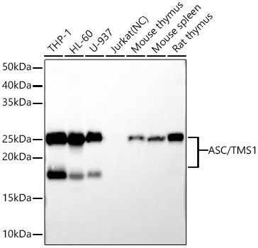
Western blot analysis of various lysates using ASC/TMS1 Rabbit mAb (A22046)at 1:12000 dilution incubated at room temperature for 1.5 hours.
Secondary antibody: HRP-conjugated Goat anti-Rabbit IgG (H+L) (AS014) at 1:10000 dilution.
Lysates/proteins: 25 μg per lane.
Blocking buffer: 3% nonfat dry milk in TBST.
Detection: ECL Basic Kit (RM00020).
Negative control (NC): Jurkat
Exposure time: 10s.
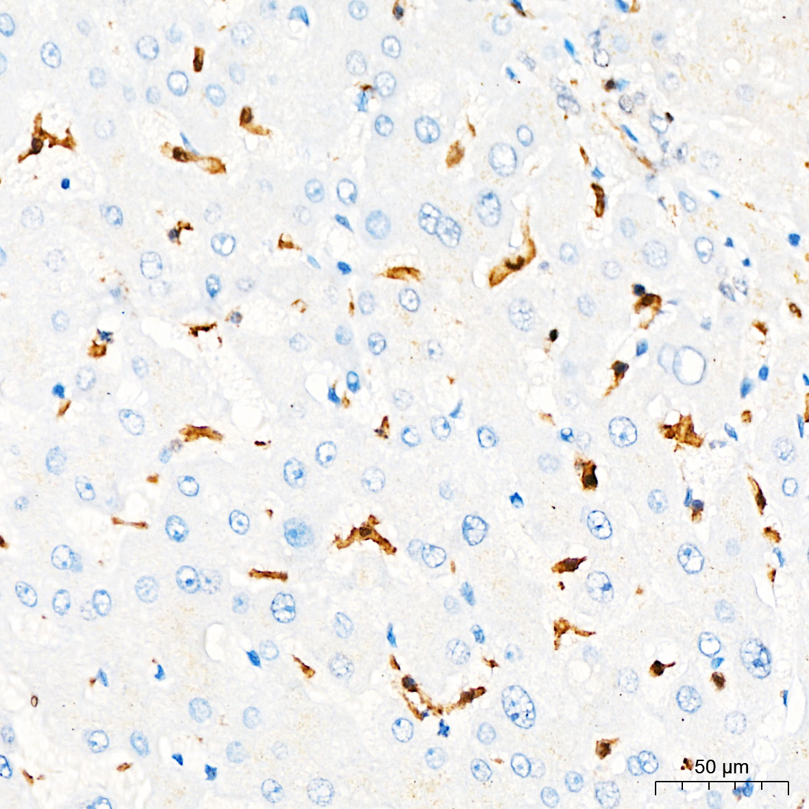
Immunohistochemistry analysis of paraffin-embedded Human liver tissue using ASC/TMS1 Rabbit mAb (A22046) at a dilution of 1:200 (40x lens). High pressure antigen retrieval performed with 0.01M Citrate buffer (pH 6.0) prior to IHC staining.
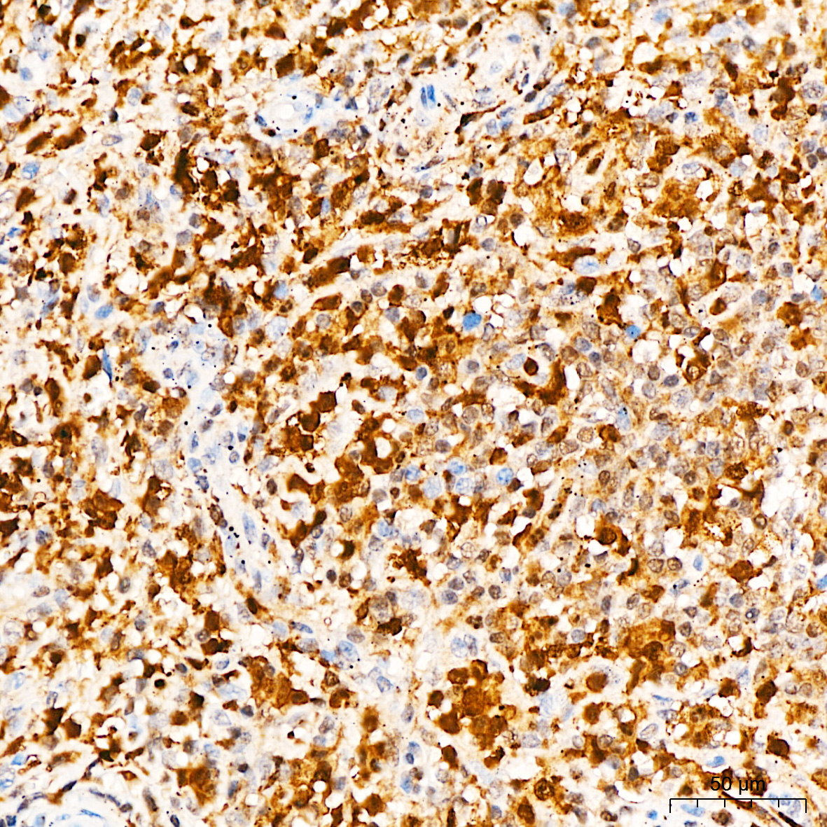
Immunohistochemistry analysis of paraffin-embedded Human spleen tissue using ASC/TMS1 Rabbit mAb (A22046) at a dilution of 1:200 (40x lens). High pressure antigen retrieval performed with 0.01M Citrate buffer (pH 6.0) prior to IHC staining.
GSDMD (Full length+C terminal) Rabbit pAb
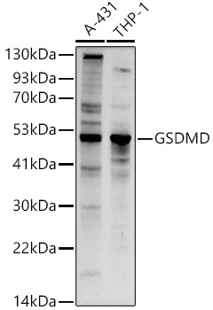
Western blot analysis of various lysates using GSDMD Rabbit pAb (A17308) at 1:1000 dilution.
Secondary antibody: HRP-conjugated Goat anti-Rabbit IgG (H+L) (AS014) at 1:10000 dilution.
Lysates/proteins: 25μg per lane.
Blocking buffer: 3% nonfat dry milk in TBST.
Detection: ECL Basic Kit (RM00020).
Exposure time: 90s.
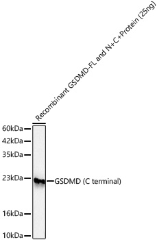
Western blot analysis of recombinant GSDMD-FL and N+C Protein using GSDMD Rabbit pAb (A17308) at 1:1000 dilution incubated overnight at 4℃.
Secondary antibody: HRP-conjugated Goat anti-Rabbit IgG (H+L) (AS014) at 1:10000 dilution.
Lysates/proteins: 25ng per lane.
Blocking buffer: 3% nonfat dry milk in TBST.
Detection: ECL Basic Kit (RM00020).
Exposure time: 60s.
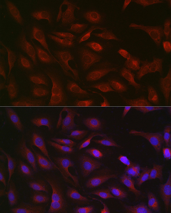
Immunofluorescence analysis of U2OS cells using GSDMD Rabbit pAb (A17308) at dilution of 1:200 (40x lens). Secondary antibody: Cy3-conjugated Goat anti-Rabbit IgG (H+L) (AS007) at 1:500 dilution. Blue: DAPI for nuclear staining.
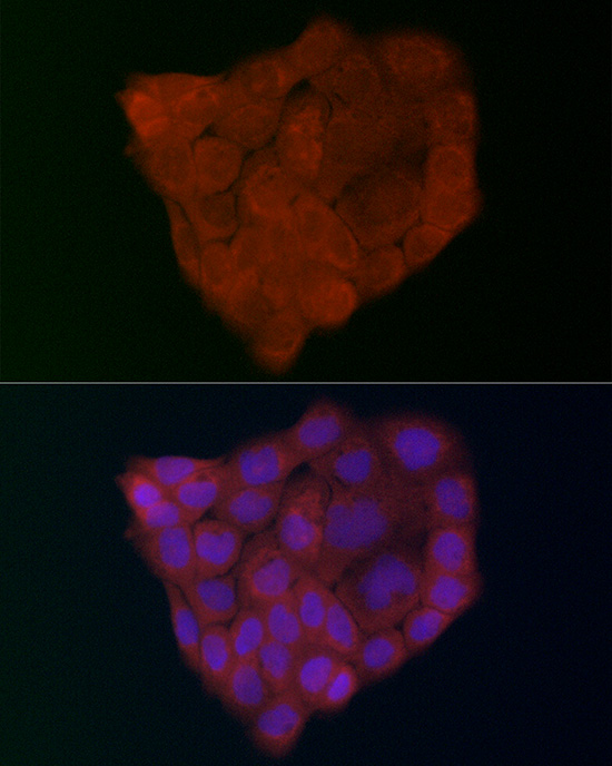
Immunofluorescence analysis of A-431 cells using GSDMD (Full Length+N terminal) Rabbit pAb (A17308) at dilution of 1:200 (40x lens). Secondary antibody: Cy3-conjugated Goat anti-Rabbit IgG (H+L) (AS007) at 1:500 dilution. Blue: DAPI for nuclear staining.
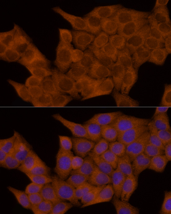
Immunofluorescence analysis of HeLa cells using GSDMD Rabbit pAb (A17308) at dilution of 1:100 (40x lens). Secondary antibody: Cy3-conjugated Goat anti-Rabbit IgG (H+L) (AS007) at 1:500 dilution. Blue: DAPI for nuclear staining.
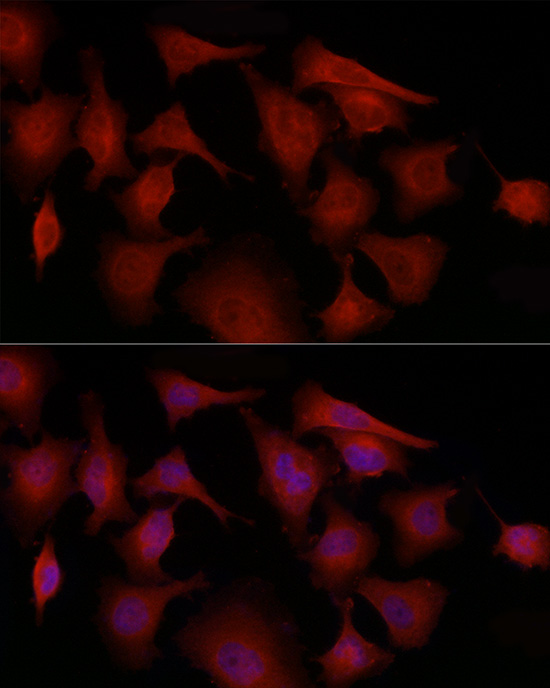
Immunofluorescence analysis of PC-3 cells using GSDMD Rabbit pAb (A17308) at dilution of 1:100 (40x lens). Secondary antibody: Cy3-conjugated Goat anti-Rabbit IgG (H+L) (AS007) at 1:500 dilution. Blue: DAPI for nuclear staining.
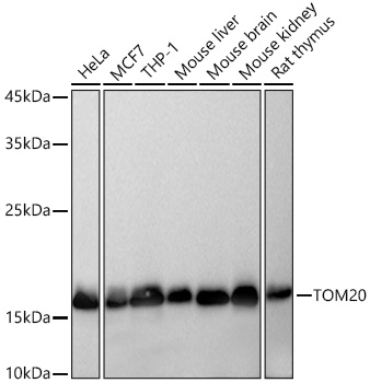
Western blot analysis of various lysates using TOM20 Rabbit mAb (A19403) at 1:5000 dilution incubated overnight at 4℃.
Secondary antibody: HRP-conjugated Goat anti-Rabbit IgG (H+L) (AS014) at 1:10000 dilution.
Lysates/proteins: 25 μg per lane.
Blocking buffer: 3% nonfat dry milk in TBST.
Detection: ECL Basic Kit (RM00020).
Exposure time: 10s.
Western blot analysis of lysates from 293T cells using TOM20 Rabbit mAb (A19403) at 1:5000 dilution incubated overnight at 4℃.
Secondary antibody: HRP-conjugated Goat anti-Rabbit IgG (H+L) (AS014) at 1:10000 dilution.
Lysates/proteins: 25 μg per lane.
Blocking buffer: 3% nonfat dry milk in TBST.
Detection: ECL Basic Kit (RM00020).
Exposure time: 10s.
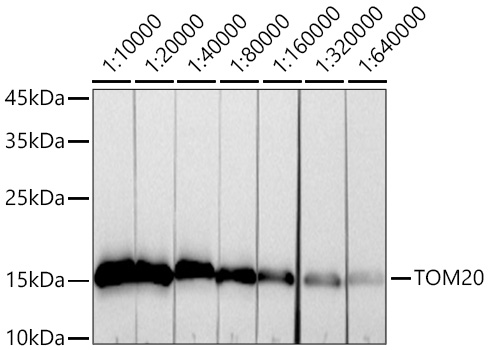
Western blot analysis of lysates from HeLa cells using TOM20 Rabbit mAb (A19403) at 1:10000-1:640000 dilution incubated overnight at 4℃.
Secondary antibody: HRP-conjugated Goat anti-Rabbit IgG (H+L) (AS014) at 1:10000 dilution.
Lysates/proteins: 25 μg per lane.
Blocking buffer: 3% nonfat dry milk in TBST.
Detection: ECL Basic Kit (RM00020).
Exposure time: 30s.
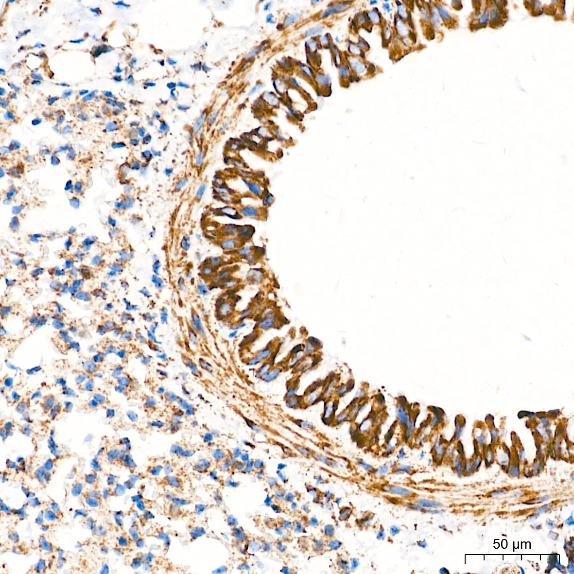
Immunohistochemistry analysis of paraffin-embedded Mouse lung tissue using TOM20 Rabbit mAb (A19403) at a dilution of 1:5000 (40x lens). High pressure antigen retrieval performed with 0.01M Tris-EDTA Buffer (pH 9.0) prior to IHC staining.
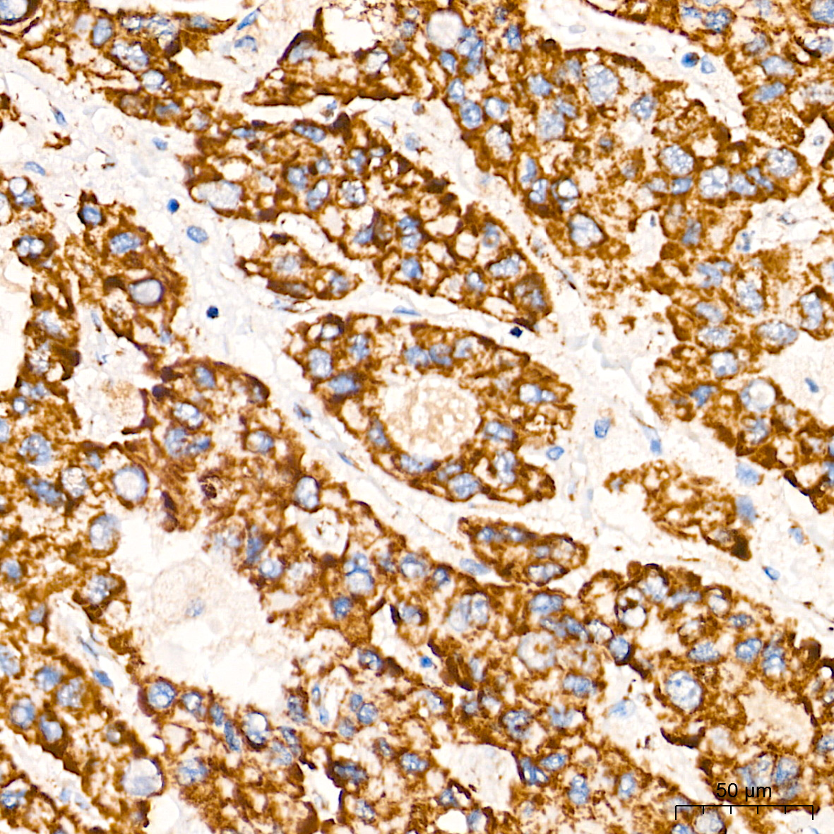
Immunohistochemistry analysis of paraffin-embedded Human liver cancer tissue using TOM20 Rabbit mAb (A19403) at a dilution of 1:5000 (40x lens). High pressure antigen retrieval performed with 0.01M Tris-EDTA Buffer (pH 9.0) prior to IHC staining.
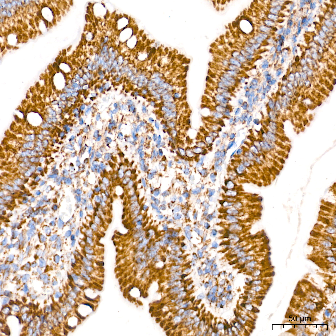
Immunohistochemistry analysis of paraffin-embedded Rat intestine tissue using TOM20 Rabbit mAb (A19403) at a dilution of 1:5000 (40x lens). High pressure antigen retrieval performed with 0.01M Tris-EDTA Buffer (pH 9.0) prior to IHC staining.
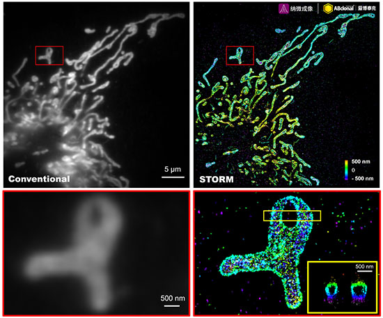
The STORM super-resolution (SR) imaging of U-2 OS cells using TOM20 Rabbit mAb (A19403, ABclonal) at dilution of 1:100 with 3% paraformaldehyde (PFA) +0.1% glutaraldehyde (GA) fixation. The immunostaining was performed by Full Automatic Immunofluorescence Workflow System (Workflow Ultra300, Nano-Micro imaging, China). Image was performed with Single-Molecule Localization Super-Resolution Microscopy (STORM Ultra300, Nano-Micro imaging, China). We acknowledge Ningbo Nano-Micro imaging Biotechnology Co., Ltd. (宁波纳微成像生物科技有限公司) in SR image processing and kindly providing this image.
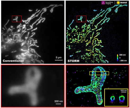
The STORM super-resolution (SR) imaging of U-2 OS cells using TOM20 Rabbit mAb (A19403, ABclonal) at dilution of 1:200 with 3% paraformaldehyde (PFA) +0.1% glutaraldehyde (GA) fixation. The immunostaining was performed by Full Automatic Immunofluorescence Workflow System (Workflow Ultra300, Nano-Micro imaging, China). Image was performed with Single-Molecule Localization Super-Resolution Microscopy (STORM Ultra300, Nano-Micro imaging, China). We acknowledge Nano-Micro imaging Biotechnology Co., Ltd. in SR image processing and kindly providing this image.
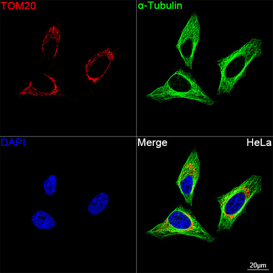
Confocal imaging of HeLa cells using TOM20 Rabbit mAb (A19403, dilution 1:2000) followed by a further incubation with Cy3 Goat Anti-Rabbit IgG (H+L) (AS007, dilution 1:500) (Red). The cells were counterstained with α-Tubulin Mouse mAb (AC012, dilution 1:400) followed by incubation with ABflo® 488-conjugated Goat Anti-Mouse IgG (H+L) Ab (AS076, dilution 1:500) (Green). DAPI was used for nuclear staining (Blue). Objective: 100x.
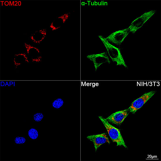
Confocal imaging of NIH/3T3 cells using TOM20 Rabbit mAb (A19403, dilution 1:2000) followed by a further incubation with Cy3 Goat Anti-Rabbit IgG (H+L) (AS007, dilution 1:500) (Red). The cells were counterstained with α-Tubulin Mouse mAb (AC012, dilution 1:400) followed by incubation with ABflo® 488-conjugated Goat Anti-Mouse IgG (H+L) Ab (AS076, dilution 1:500) (Green). DAPI was used for nuclear staining (Blue). Objective: 100x.
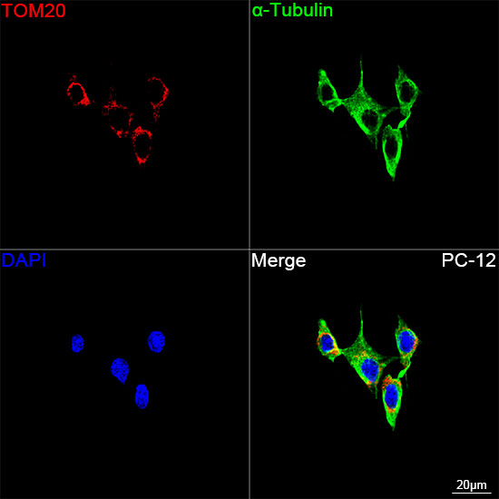
Confocal imaging of PC-12 cells using TOM20 Rabbit mAb (A19403, dilution 1:2000) followed by a further incubation with Cy3 Goat Anti-Rabbit IgG (H+L) (AS007, dilution 1:500) (Red). The cells were counterstained with α-Tubulin Mouse mAb (AC012, dilution 1:400) followed by incubation with ABflo® 488-conjugated Goat Anti-Mouse IgG (H+L) Ab (AS076, dilution 1:500) (Green). DAPI was used for nuclear staining (Blue). Objective: 100x.
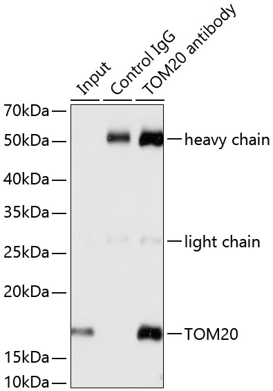
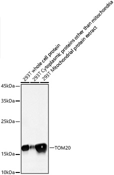
Immunoprecipitation analysis of 200 μg extracts from HeLa cells using 3 μg TOM20 antibody (A19403). Western blot was performed from the immunoprecipitate using TOM20 antibody (A19403) at a dilution of 1:1000.
| 分类 |
|
|||||||||||||
|---|---|---|---|---|---|---|---|---|---|---|---|---|---|---|
|
炎症小体成分 (感受器) |
|
|||||||||||||
|
炎症小体成分 (接头分子) |
|
|||||||||||||
|
炎症小体成分 (促炎caspase) |
|
|||||||||||||
| 促炎细胞因子 |
|
|||||||||||||
| 细胞焦亡效应分子 |
|
|||||||||||||