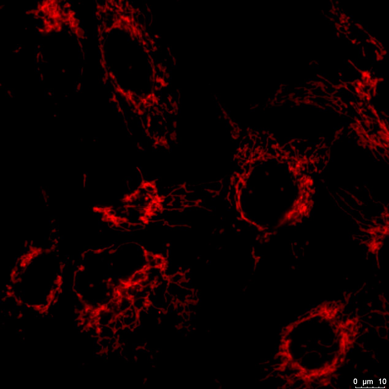
| WB (Western Blot) | Human, Mouse, Rat, Pig, Other |
|---|---|
| IP (Immunoprecipitation) | Mouse |
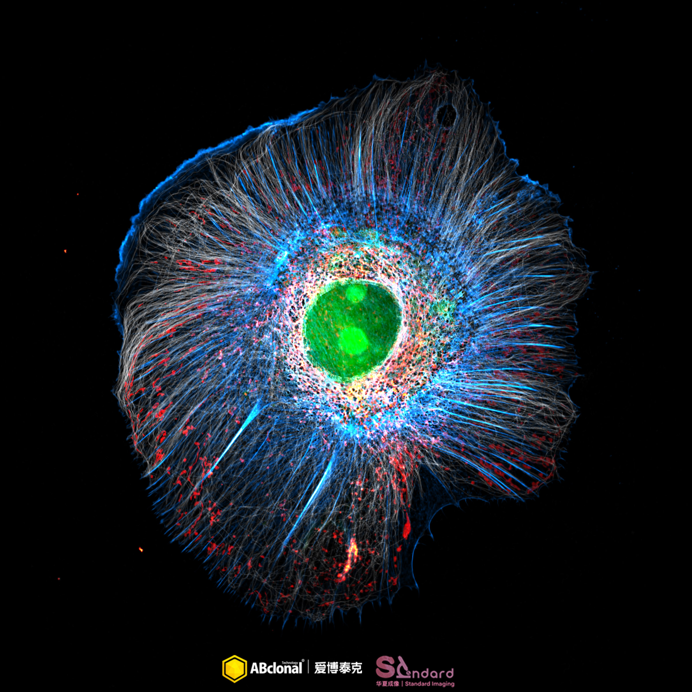
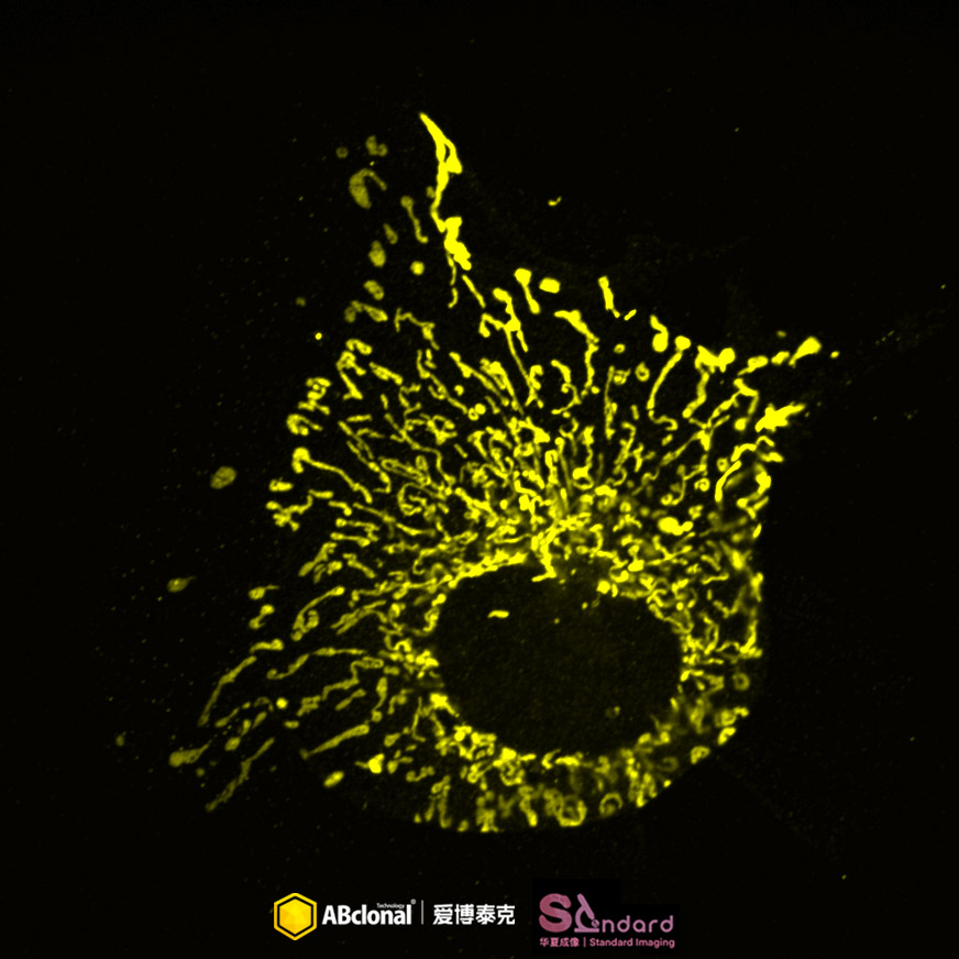
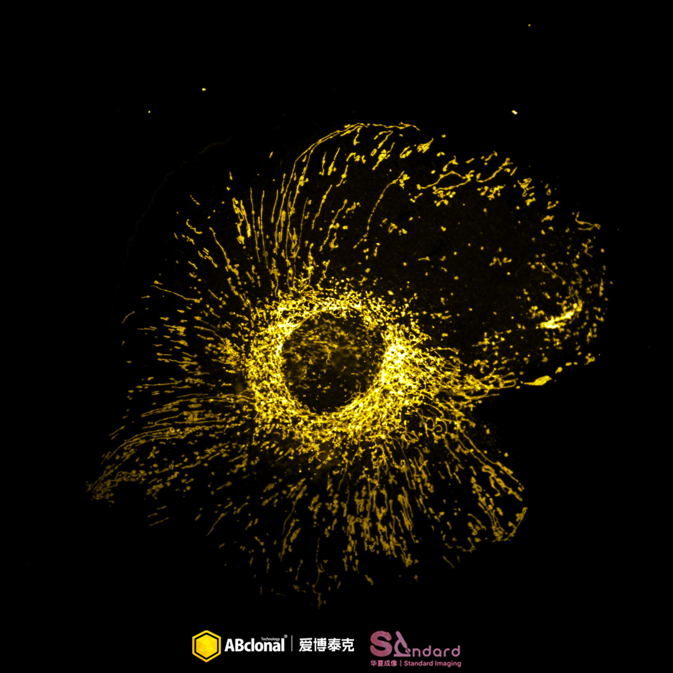
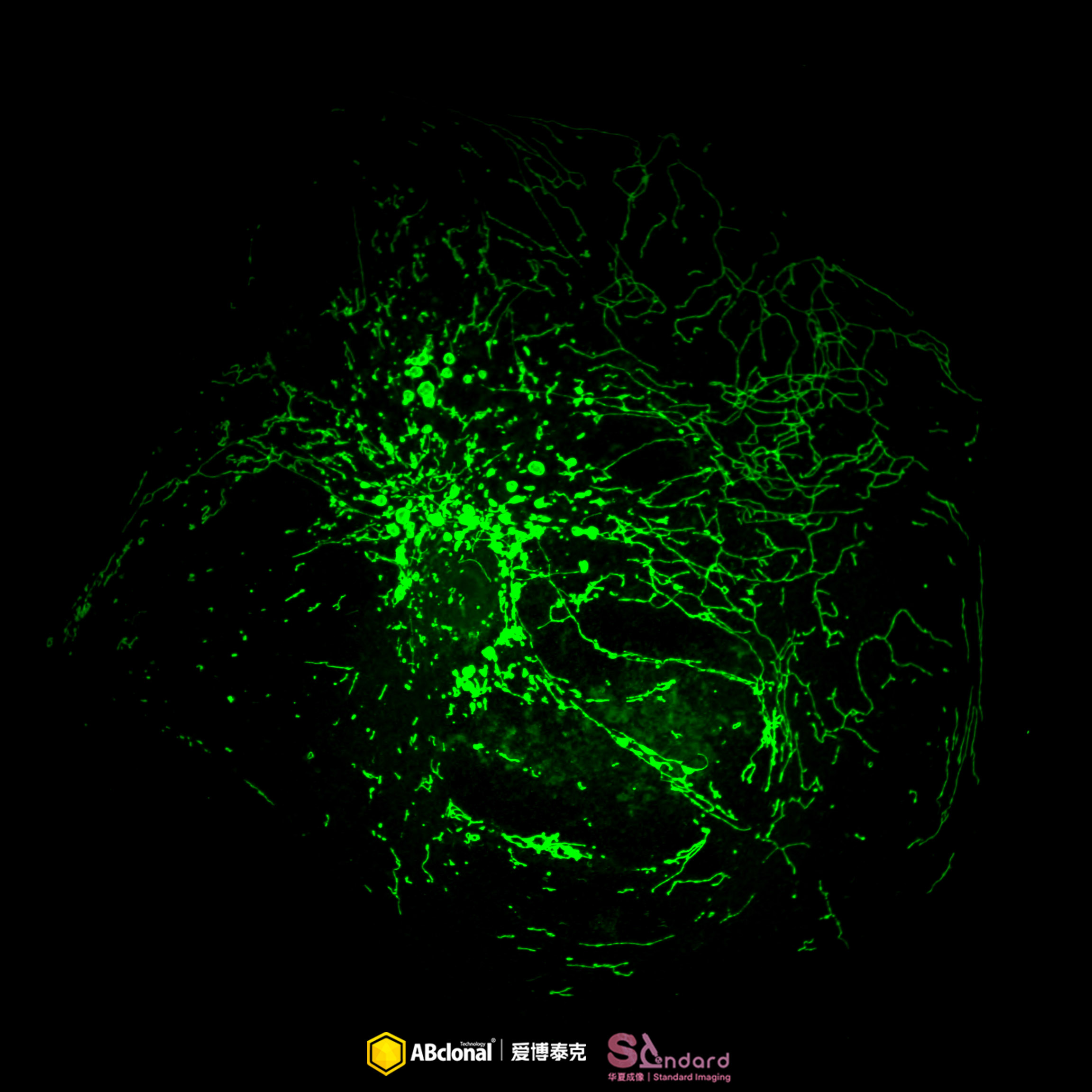
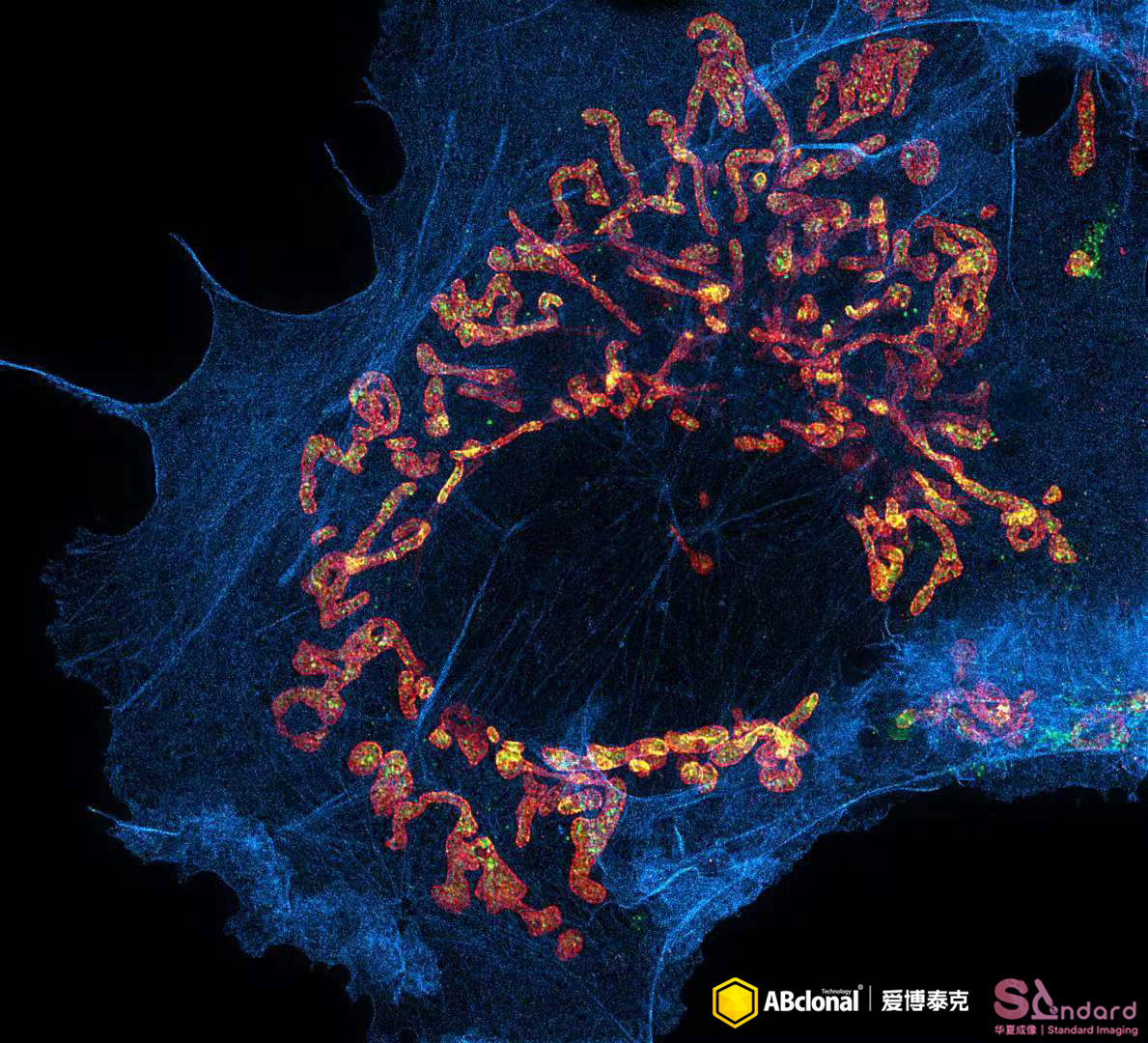
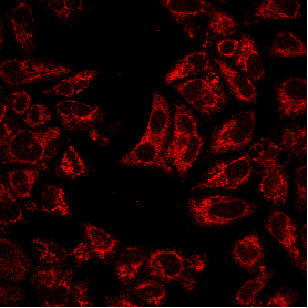
| IF (Immunofluorescence) | Human, Mouse, Rat, Other |
| FC (Flow Cytometry) | Human |
| IHC (Immunohistochemistry) | Human, Mouse, Rat, Other |
请输入产品标签上的lot号,例如4000000001


Publishing research using A19403? Please let us know so that we can cite the reference in this datasheet.
首先,一般抗体不推荐客户回收利用,抗体使用之后缓冲体系已经发生改变,不同客户在回收抗体的保存条件上也会有差异,所以抗体回收使用效果无法保证。另外,ABclonal公司也做过一批抗体回收验证测试,测试结果显示不同抗体可回收次数不同,一般效价越高的抗体,可重复使用的次数越多,客户可根据实验情况来确定。
注:我们将孵育完毕后剩余的抗体回收到离心管中置于4℃保存,效价高的抗体可至少保存1周,至少重复利用3次。
武汉爱博泰克生物(ABclonal)科技有限公司是国产品牌,她成立于2011年,公司依托ABclonal美国波士顿抗体与蛋白研发中心、中国光谷生物城(武汉)抗体生产基地以及上海张江分子酶研发中心,凝聚了十余位来自哈佛大学、麻省理工、复旦大学、上海交大、中科院生化细胞所和武汉大学的全球知名分子和免疫学方面博士,组成我们的科学家团队,通过聚焦抗体与酶核心技术,致力于打破国际技术的垄断,将公司打造成为科研工具和诊断原料的国内领导品牌,乃至弯道超越国际巨头。 我们拥有包括兔多克隆抗体、小鼠单克隆抗体、兔单克隆抗体的生产研发平台,同时也有包括WB,IHC,IF,IP,CHIP在内的检测平台,我们对每一支自产的抗体进行了严格的检测。当然,我们部分直销地区也可以帮客户代购进口品牌的产品。同时也有抗体定制服务。ABclonal抗体优势:1,严自检,保质量;2产品多,指标全;3,价格低,货期短。注:ABclonal抗体价格体系详情见附件
ABclonal抗体成分在平时工作当中,常会有客户咨询我们的抗体用的什么buffer进行保存,一般来说,我们的buffer的成分是:PBS含0.03%的proclin300、0.05%牛血清白蛋白、50%甘油;也有一些是PBS含0.03%的proclin300,50%甘油。防腐剂 Proclin 300活性成分主要是2-甲基-4-异噻唑啉-3-酮(MCI)和5-氯-2-甲基-4-异噻唑啉-3-酮(CMCI)。ProClin生物灭活剂能够迅速穿透细胞膜,抑制对细胞呼吸至关重要的特定酶,因此一接触微生物有机体就会立即抑制细胞活性。ProClin的多个特定毒性位点可以防止微生物产生高水平的耐药性。

_WB_01.jpg?t=1756896765)