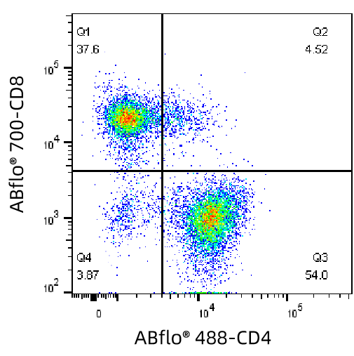
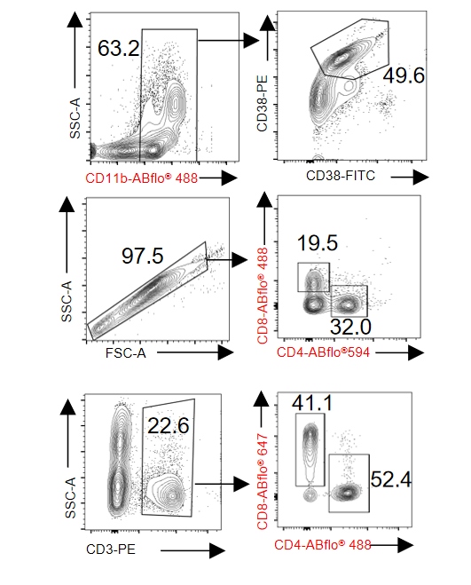
Flow cytometry analysis of mouse spleen tissue using ABflo® 700 Rabbit anti-Mouse CD8a mAb (A25944) and ABflo® 488 Rabbit anti-Mouse CD4 mAb (A24720) at 1:100 dilution. clearly classified positive and negative cells.
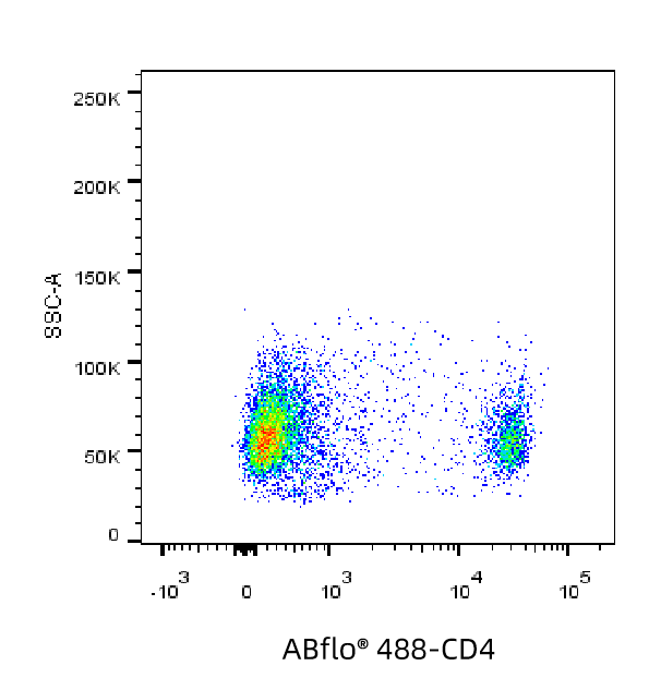
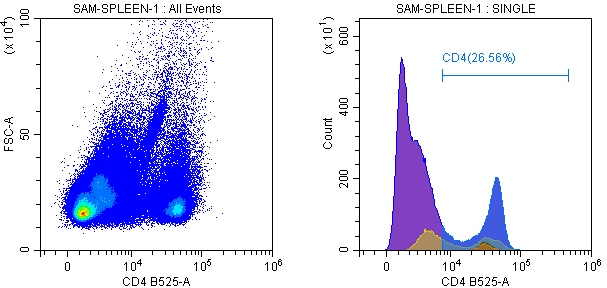
Flow cytometry analysis of mouse T lymphocyte using ABflo® 488 Rabbit anti-Mouse CD4 mAb (A24720) at 1:20 dilution. clearly classified positive and negative cells.
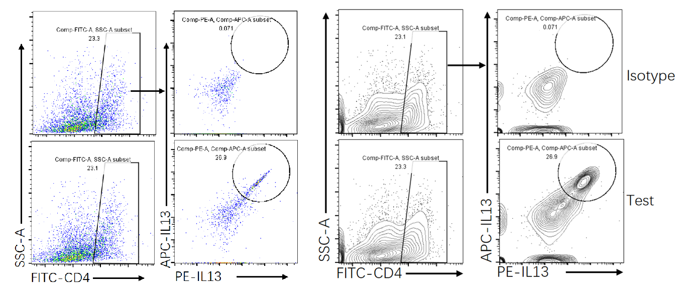
Flow cytometry analysis of T cells in the tumor microenvironment of mouse lungs using APC Rabbit anti-Mouse IL-13 mAb (A26688) , PE Rabbit anti-Mouse IL-13 mAb (A26687) and ABflo® 488 Rabbit anti-Mouse CD4 mAb (A24720) clearly classified positive and negative cells.
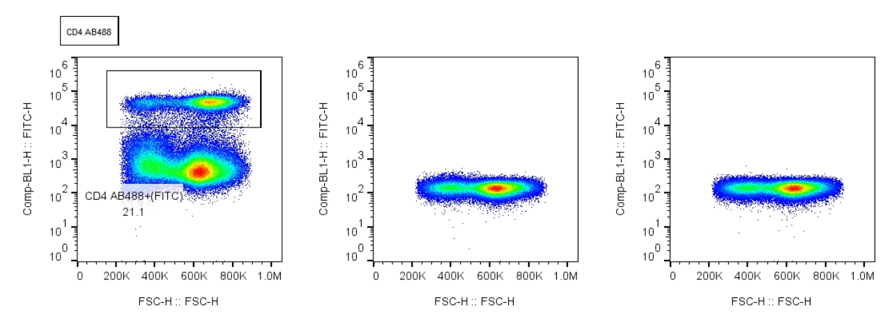
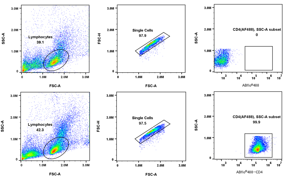
The figure shows the gating strategy for a blank tube (top) and an ABclonal antibody ABflo®488 Rabbit anti-Mouse CD4 mAb single-staining tube (bottom). The positive signal transition is obvious.
Predicted to enable several functions, including interleukin-16 binding activity; interleukin-16 receptor activity; and protein homodimerization activity. Involved in helper T cell enhancement of adaptive immune response. Acts upstream of or within several processes, including defense response to Gram-negative bacterium; positive regulation of calcium-mediated signaling; and positive regulation of peptidyl-tyrosine phosphorylation. Located in endoplasmic reticulum; external side of plasma membrane; and membrane raft. Is expressed in several structures, including alimentary system; brain; genitourinary system; hemolymphoid system; and liver and biliary system. Used to study type 1 diabetes mellitus. Human ortholog(s) of this gene implicated in immunodeficiency 79. Orthologous to human CD4 (CD4 molecule).