利用敲除、敲低技术,将目标基因敲除或敲低以验证抗体特异性。当看到 KO/KD验证标记时,您可以相信该抗体不仅在推荐的应用和种属中得到了验证,而且其特异性也已经通过我们内部的KO/KD验证方法得到了确认。
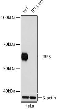
Western blot analysis of lysates from wild type (WT) and IRF3 knockout (KO) HeLa cells, using [KO Validated] IRF3 Rabbit mAb (A19717) at 1:1000 dilution.
Secondary antibody: HRP Goat Anti-Rabbit IgG (H+L) (AS014) at 1:10000 dilution.
Lysates/proteins: 25μg per lane.
Blocking buffer: 3% nonfat dry milk in TBST.
Detection: ECL Basic Kit (RM00020).
Exposure time: 3min.
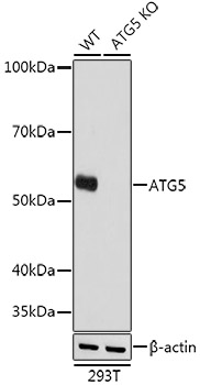
Western blot analysis of lysates from wild type (WT) and ATG5 knockout (KO) 293T cells, using [KO Validated] ATG5 Rabbit mAb (A19677) at 1:1000 dilution.
Secondary antibody: HRP Goat Anti-Rabbit IgG (H+L) (AS014) at 1:10000 dilution.
Lysates/proteins: 25μg per lane.
Blocking buffer: 3% nonfat dry milk in TBST.
Detection: ECL Basic Kit (RM00020).
Exposure time: 1s.
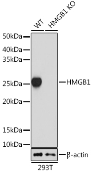
Western blot analysis of extracts from wild type (WT) and HMGB1 knockout (KO) 293T cells, using HMGB1 antibody (A19529) at 1:1000 dilution.
Secondary antibody: HRP Goat Anti-Rabbit IgG (H+L) (AS014) at 1:10000 dilution.
Lysates/proteins: 25μg per lane.
Blocking buffer: 3% nonfat dry milk in TBST.
Detection: ECL Basic Kit (RM00020).
Exposure time: 5s.
过表达目标蛋白以验证抗体的特异性。在表现出弱内源性表达或无内源性表达的细胞系中,过表达靶蛋白可以给出抗体特异性和敏感性的问题。组织或细胞裂解物中预期大小或定位的干净信号,已知含有目的蛋白质,结合过表达系统中的阳性结果,是特异性验证的良好指标。
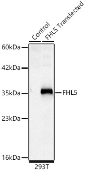
Western blot analysis of lysates from wild type (WT) and 293T cells transfected with FHL5 using FHL5 Rabbit mAb(A25133) at 1:4000 dilution.
Secondary antibody: HRP Goat Anti-Rabbit IgG (H+L) (AS014) at 1:10000 dilution.
Lysates/proteins: 25 μg per lane.
Blocking buffer: 3% nonfat dry milk in TBST.
Detection: ECL Basic Kit (RM00020).
Exposure time: 5s.
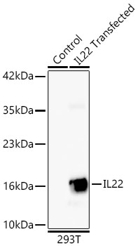
Western blot analysis of extracts of normal 293T cells and 293T transfected with IL22 using IL22 Rabbit mAb (A24612) at 1:1000 dilution.
Secondary antibody: HRP Goat Anti-Rabbit IgG (H+L) (AS014) at 1:10000 dilution.
Lysates/proteins: 25ug per lane.
Blocking buffer: 3% nonfat dry milk in TBST.
Detection: ECL Basic Kit (RM00020).
Exposure time: 45s.
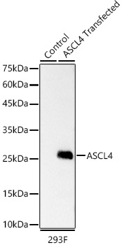
Western blot analysis of lysates from wild type (WT) and 293F cells transfected with ASCL4 using ASCL4 Rabbit mAb (A23548) at 1:5000 dilution.
Secondary antibody: HRP Goat Anti-Rabbit IgG (H+L) (AS014) at 1:10000 dilution.
Lysates/proteins: 25 μg per lane.
Blocking buffer: 3% nonfat dry milk in TBST.
Detection: ECL Basic Kit (RM00020).
Exposure time: 0.3s.
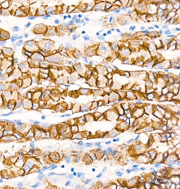
同靶点多表位抗体验证是一种强大的抗体验证策略,使用针对同一靶标上不同、不重叠表位的两种或多种抗体,以产生直接可比的免疫染色数据。通过平行地用多种抗体检测相同的样品,可以相对快速直观地了解抗体特异性。这两种抗体A22850(ARC57093)、A20798(ARC51012)可检测人胃组织E-Cadherin上的独立、独特表位。使用两种抗体获得的相似染色模式有助于确认染色特异性。
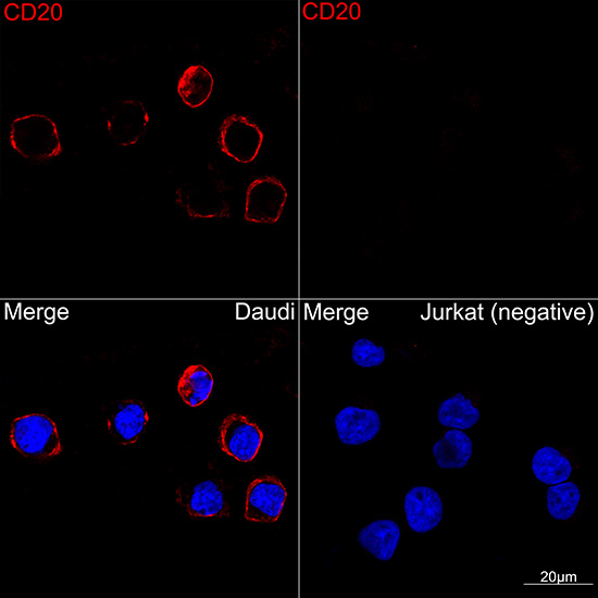
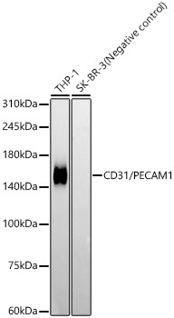
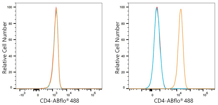
蛋白质在部分细胞或组织类型中表达,而在其他细胞或组织类型中则不表达。通过不同的应用(免疫印迹、免疫荧光、流式细胞术等)分析这些蛋白在不同的细胞或组织类型之中的差异表达情况,可确定抗体靶向特异性。
Flow cytometry: 1X10^6 PC-3 cells (negative control,left) and THP-1 cells (right) were surface-stained with ABflo® 488 Rabbit anti-Human CD4 mAb (A22773,5 μl/Test,orange line) or ABflo® 488 Rabbit IgG isotype control (A22069,5 μl/Test,blue line). Non-fluorescently stained cells were used as blank control (red line).
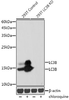
以在特定的生理病理条件下,靶点蛋白的生物学变化作为基础进行定性验证,如细胞处理后检测下游信号。
Western blot analysis of lysates from wild type(WT) and LC3B knockdown (KD) 293T cells, using [KD Validated] LC3B Rabbit mAb (A19665) at 1:1000 dilution.wild type(WT) and LC3B knockdown (KD) 293T cells were treated by Chloroquine (50 μM) at 37℃ for 20 hours.
Secondary antibody: HRP Goat Anti-Rabbit IgG (H+L) (AS014) at 1:10000 dilution.
Lysates/proteins: 25μg per lane.
Blocking buffer: 3% nonfat dry milk in TBST.
Detection: ECL Basic Kit (RM00020).
Exposure time: 30s.
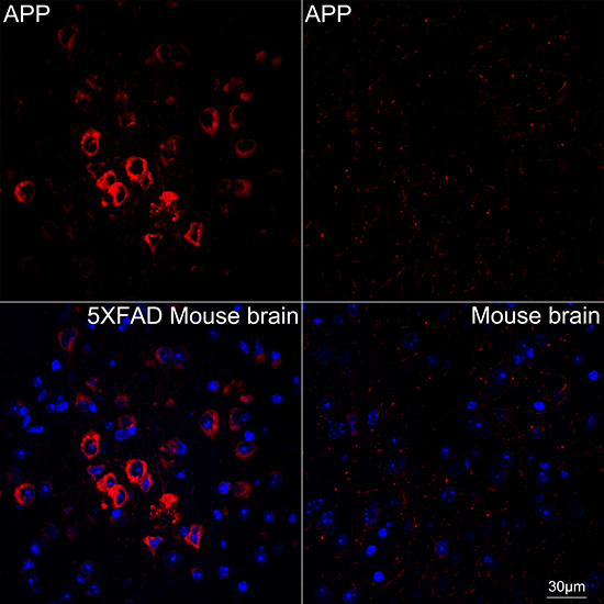
Confocal imaging of paraffin-embedded 5XFAD Mouse brain and Mouse brain using [KO Validated] APP Rabbit mAb (A17911, dilution 1:200) followed by a further incubation with Cy3 Goat Anti-Rabbit IgG (H+L) (AS007, dilution 1:500) (Red). DAPI was used for nuclear staining (Blue). Objective: 40x.
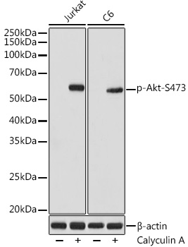
Western blot analysis of various lysates using Phospho-Akt-S473 Rabbit mAb (AP1208) at 1:1000 dilution. Both Jurkat and C6 cells were treated by Calyculin A (100 nM) at 37℃ for 30 minutes after serum-starvation overnight.
Secondary antibody: HRP-conjugated Goat anti-Rabbit IgG (H+L) (AS014) at 1:10000 dilution.
Lysates/proteins: 25μg per lane.
Blocking buffer: 3% nonfat dry milk in TBST.
Detection: ECL Basic Kit (RM00020).
Exposure time: 1s.
抗体多种方法联合检测验证抗体特异性,如组蛋白及组蛋白修饰通过不同的应用(免疫印迹、免疫组化分析、ChIP-seq 等)分析蛋白表达的特异性。
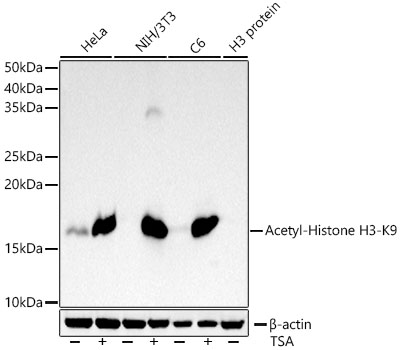
Western blot analysis of various lysates using Acetyl-Histone H3-K9 Rabbit mAb (A21107) at1:1000 dilution. HeLa and NIH/3T3 and C6 cells were treated by TSA (1 uM) at 37℃ for 18 hours.
Secondary antibody: HRP-conjugated Goat anti-Rabbit IgG (H+L) (AS014) at1:10000 dilution.
Lysates/proteins: 25μg per lane.
Blocking buffer: 3% nonfat dry milk in TBST.
Detection: ECL Basic Kit (RM00020).
Exposure time: 10s.
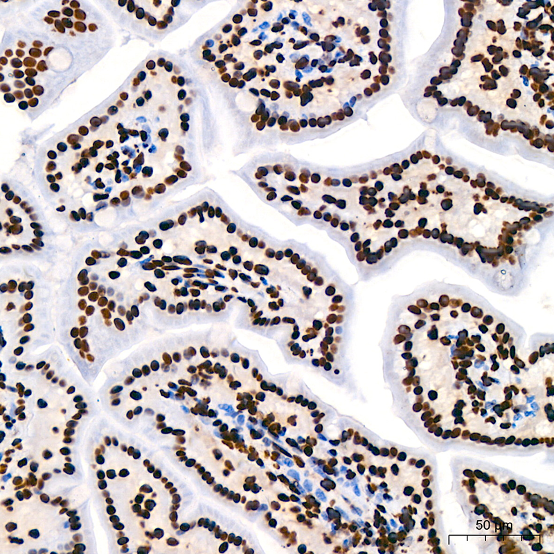
Immunohistochemistry analysis of paraffin-embedded Mouse intestin using Acetyl-Histone H3-K9 Rabbit mAb (A21107) at dilution of 1:200 (40x lens). High pressure antigen retrieval performed with 0.01M Citrate Bufferr (pH 6.0) prior to IHC staining.
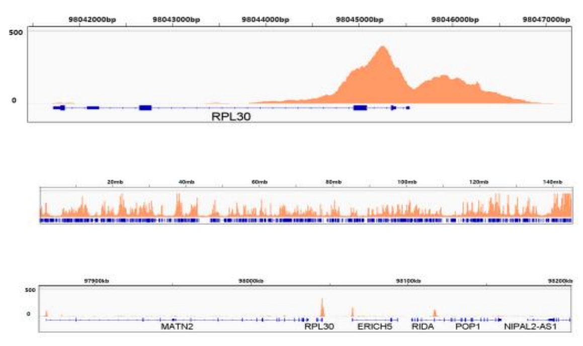
Chromatin immunoprecipitations were performed with cross-linked chromatin from 293F cells and Acetyl-Histone H3-K9 (A21107). The ChIP sequencing results indicate the enrichment pattern of Acetyl-Histone H3-K9 in selected genomic region and representative gene loci (RPL30), as shown in figure.