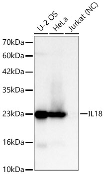
Western blot analysis of various lysates, using IL18 Rabbit mAb (A23076) at 1:7000 dilution.
Secondary antibody: HRP Goat Anti-Rabbit IgG (H+L) (AS014) at 1:10000 dilution.
Lysates/proteins: 25μg per lane.
Blocking buffer: 3% nonfat dry milk in TBST.
Detection: ECL Basic Kit (RM00020).
Exposure time: 30s.
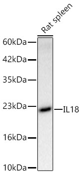
Western blot analysis of lysates from Rat spleen using IL18 Rabbit mAb (A23076) at 1:1000 dilution incubated overnight at 4℃.
Secondary antibody: HRP-conjugated Goat anti-Rabbit IgG (H+L)(AS014) at 1:10000 dilution.
Lysates/proteins: 25 μg per lane.
Blocking buffer: 3% nonfat dry milk in TBST.
Detection: ECL Basic Kit (RM00020)
Exposure time: 45 s.

Immunohistochemistry analysis of IL18 in paraffin-embedded human esophagus tissue using IL18 Rabbit mAb (A23076) at a dilution of 1:1000 (40x lens).High pressure antigen retrieval was performed with 0.01 M citrate buffer (pH 6.0) prior to IHC staining.

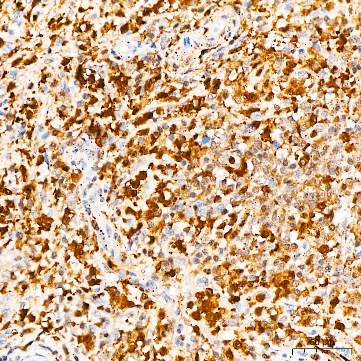
Immunohistochemistry analysis of IL18 in paraffin-embedded human tonsil tissue using IL18 Rabbit mAb (A23076) at a dilution of 1:1000 (40x lens).High pressure antigen retrieval was performed with 0.01 M citrate buffer (pH 6.0) prior to IHC staining.
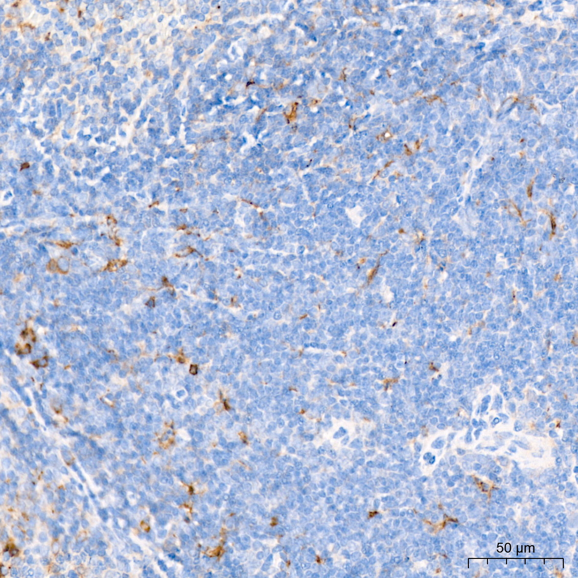
Immunohistochemistry analysis of IL18 in paraffin-embedded mouse spleen tissue using IL18 Rabbit mAb (A23076) at a dilution of 1:1000 (40x lens).High pressure antigen retrieval was performed with 0.01 M citrate buffer (pH 6.0) prior to IHC staining.
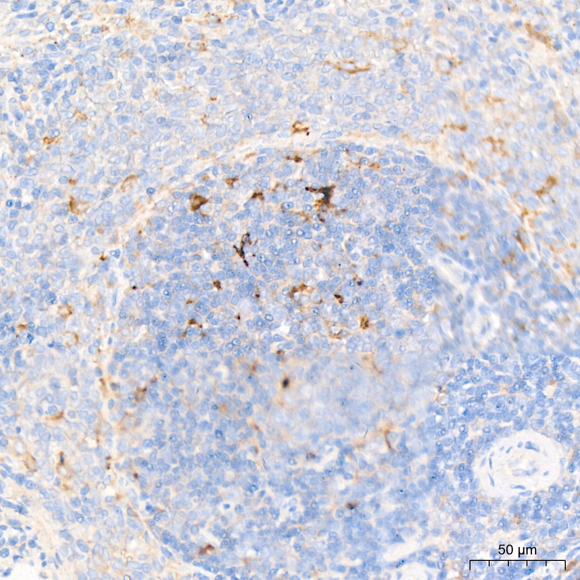
Immunohistochemistry analysis of IL18 in paraffin-embedded rat spleen tissue using IL18 Rabbit mAb (A23076) at a dilution of 1:1000 (40x lens).High pressure antigen retrieval was performed with 0.01 M citrate buffer (pH 6.0) prior to IHC staining.

Western blot analysis of various lysates, using CASP1 Rabbit pAb (A0964) at 1:1800 dilution.
Secondary antibody: HRP Goat Anti-Rabbit IgG (H+L) (AS014) at 1:10000 dilution.
Lysates/proteins: 25μg per lane.
Blocking buffer: 3% nonfat dry milk in TBST.
Detection: ECL Enhanced Kit (RM00021).
Exposure time: 60s.
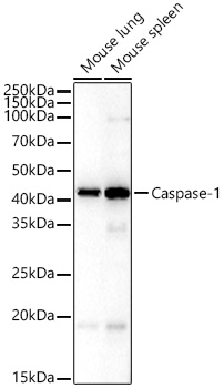
Western blot analysis of various lysates, using CASP1 Rabbit pAb (A0964) at 1:1800 dilution.
Secondary antibody: HRP Goat Anti-Rabbit IgG (H+L) (AS014) at 1:10000 dilution.
Lysates/proteins: 25μg per lane.
Blocking buffer: 3% nonfat dry milk in TBST.
Detection: ECL Basic Kit (RM00020).
Exposure time: 180s.
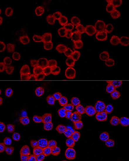
Confocal immunofluorescence analysis of Raw264.7 cells using Caspase-1 Rabbit pAb (A0964) at dilution of 1:200. Blue: DAPI for nuclear staining.

Immunofluorescence analysis of PC-12 cells using CASP1 Rabbit pAb (A0964) at dilution of 1:100 (40x lens). Secondary antibody: Cy3 Goat Anti-Rabbit IgG (H+L) (AS007) at 1:500 dilution. Blue: DAPI for nuclear staining.
_IF_01.jpg?t=1706682197)
Immunofluorescence analysis of NIH/3T3 cells using Caspase-1 Rabbit pAb (A0964) at dilution of 1:200 (40x lens). Secondary antibody: Cy3 Goat Anti-Rabbit IgG (H+L) (AS007) at 1:500 dilution. Blue: DAPI for nuclear staining.
_IF_02.jpg?t=1706682197)
Immunofluorescence analysis of U2OS cells using Caspase-1 Rabbit pAb (A0964) at dilution of 1:200 (40x lens). Secondary antibody: Cy3 Goat Anti-Rabbit IgG (H+L) (AS007) at 1:500 dilution. Blue: DAPI for nuclear staining.
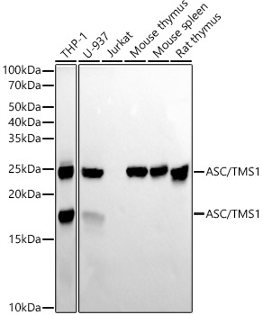
Western blot analysis of various lysates using ASC/TMS1 Rabbit mAb (A22046) at1:6000 dilution.
Secondary antibody: HRP Goat Anti-Rabbit IgG (H+L) (AS014) at 1:10000 dilution.
Lysates/proteins: 25μg per lane.
Blocking buffer: 3% nonfat dry milk in TBST.
Detection: ECL Basic Kit (RM00020).
Exposure time: 10s.
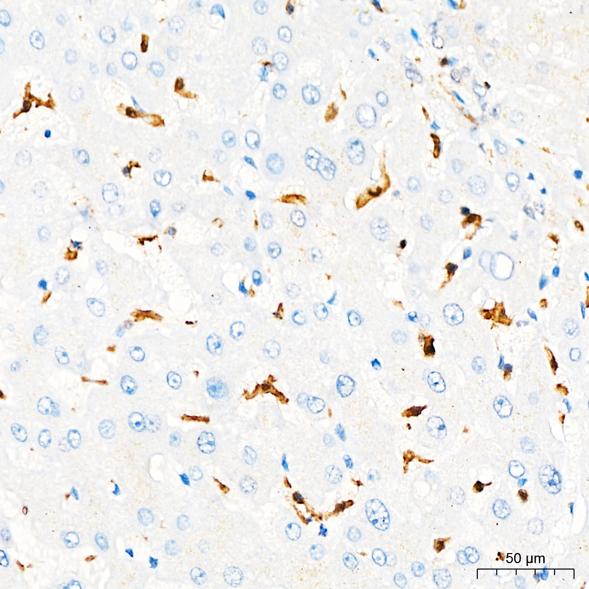
Immunohistochemistry analysis of paraffin-embedded Human liver tissue using ASC/TMS1 Rabbit mAb (A22046) at a dilution of 1:200 (40x lens). High pressure antigen retrieval performed with 0.01M Citrate Buffer(pH 6.0) prior to IHC staining.
Immunohistochemistry analysis of paraffin-embedded Human spleen tissue using ASC/TMS1 Rabbit mAb (A22046) at a dilution of 1:200 (40x lens). High pressure antigen retrieval performed with 0.01M Citrate Buffer(pH 6.0) prior to IHC staining.
GSDMD (Full length+C terminal) Rabbit mAb
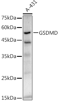
Western blot analysis of lysates from A-431 cells, using GSDMD Rabbit mAb (A20728) at 1:1000 dilution.
Secondary antibody: HRP Goat Anti-Rabbit IgG (H+L) (AS014) at 1:10000 dilution.
Lysates/proteins: 25μg per lane.
Blocking buffer: 3% nonfat dry milk in TBST.
Detection: ECL Enhanced Kit (RM00021).
Exposure time: 180s.
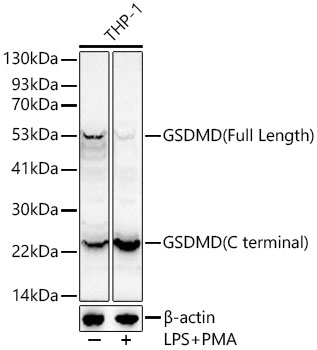
Western blot analysis of lysates from THP-1 cells, using GSDMD Rabbit mAb (A20728) at 1:1000 dilution.THP-1 cells were treated by PMA/TPA (80 nM) at 37℃ for overnight and LPS (1 μg/ml) at 37℃ for 6 hours.
Secondary antibody: HRP Goat Anti-Rabbit IgG (H+L) (AS014) at 1:10000 dilution.
Lysates/proteins: 25μg per lane.
Blocking buffer: 3% nonfat dry milk in TBST.
Detection: ECL Enhanced Kit (RM00021).
Exposure time: 60s.
| 分类 |
|
||||||||||||||||||||||||||||||||||||||||||||||||||||||
|---|---|---|---|---|---|---|---|---|---|---|---|---|---|---|---|---|---|---|---|---|---|---|---|---|---|---|---|---|---|---|---|---|---|---|---|---|---|---|---|---|---|---|---|---|---|---|---|---|---|---|---|---|---|---|---|
|
炎症小体成分 (感受器) |
|
||||||||||||||||||||||||||||||||||||||||||||||||||||||
|
炎症小体成分 (接头分子) |
|
||||||||||||||||||||||||||||||||||||||||||||||||||||||
|
炎症小体成分 (促炎caspase) |
|
||||||||||||||||||||||||||||||||||||||||||||||||||||||
| 促炎细胞因子 |
|
||||||||||||||||||||||||||||||||||||||||||||||||||||||
| 细胞焦亡效应分子 |
|
||||||||||||||||||||||||||||||||||||||||||||||||||||||